The medial pterygoid muscle functions to assist with elevation and protrusion of the mandible. What is the action of the masseter muscle.
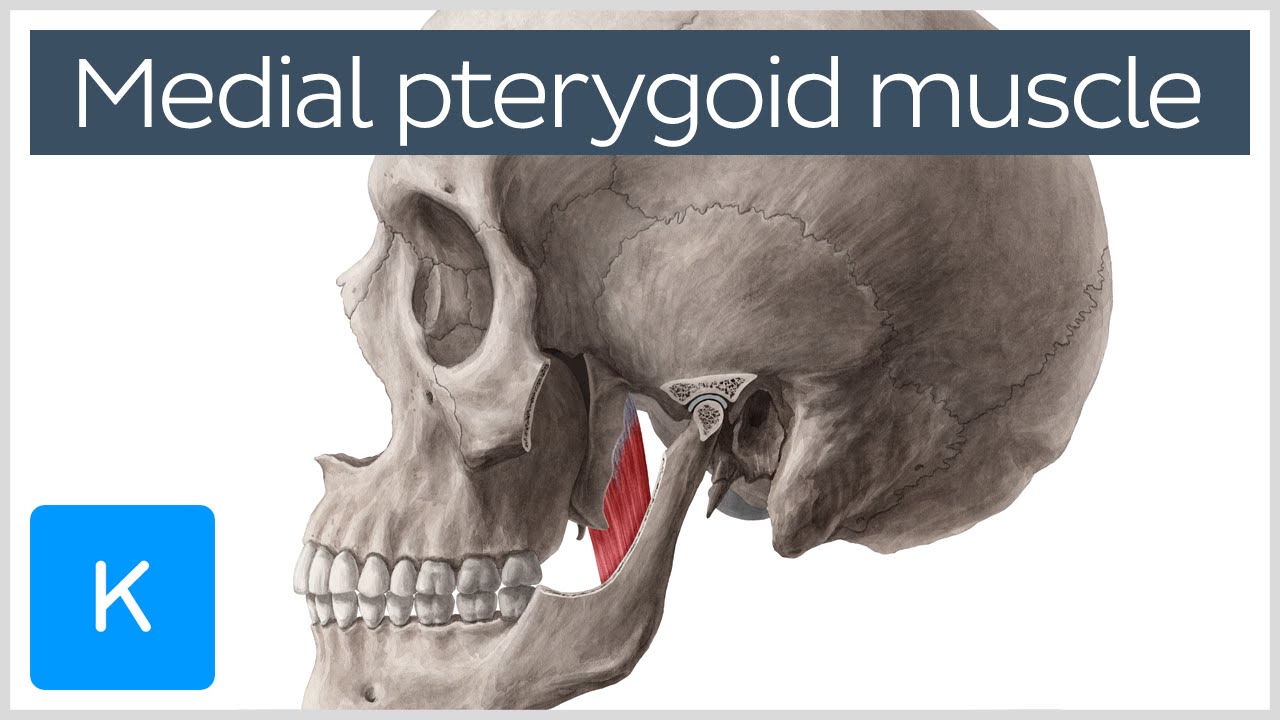
Medial Pterygoid Muscle Origin Insertion Function Nerve Supply Anatomy Kenhub Youtube
Musculus pterygoideus lateralis is a thick and short triangular-shaped muscle located in the infratemporal fossa of the skull.
. Elevation of the mandible occurs during the closing of the jaws. It can assist in protrusion of the mandible. Specifically the medial pterygoid muscle functions to.
Portable and easy to use Medial Pterygoid Muscle Action study sets help you review the information and examples you need to succeed in the time you have available. It arises from the medial surface of the lateral pterygoid plate and the grooved surface of the pyramidal process of the palatine bone. Elevates mandible closes jaw helps lateral pterygoids in moving the jaw from side to side.
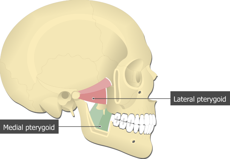
The medial pterygoid is a thick quadrilateral muscle. The deep head is the major component and is attached to the medial aspect of the lateral pterygoid plate of the sphenoid bone. Lateral Pterygoid The lateral pterygoid muscle is the primary muscle of the inferior temporal fossa.
See appendix 3-4 and see color plates. From medial surface of lateral pterygoid plate adjoining pro. Learn the facial muscles easily with these quizzes and labeled diagrams.
The medial pterygoid muscle is a thick and square shaped muscle. Raiseelevate the lower jaw. Medial pterygoid muscle consists of two heads.
Protraction of the mandible when medial and lateral pterygoid muscles contract bilaterally. The function of a muscle is usually associated with the part of the body to which the muscles attaches. The fibers of the medial pterygoid muscle run posterolaterally and insert to the medial surface of the mandibular ramus and angle.
Mandibular nerve via nerve to medial pterygoid. This muscle also contributes to the elevation of the mandible acting as a synergist to the temporalis and masseter. It is classified as one of the primary muscles of mastication.
It has two heads of origin. Protract or protrude mandible. The lateral pterygoid muscle is composed of two heads - superior and inferior.
It has a second slip of origin. Medial pterygoid is a thick quadrilateral muscle that connects the mandible with maxilla sphenoid and palatine bones. By contracting on one side the medial pterygoid pushes the mandible to the opposite side.
Since the medial pterygoid muscle attaches to the lower jaw the function of this muscle is to move the lower jaw. The lateral pterygoid muscle is a small thick muscle located on each side of the skull that assists with mastication chewing. Since the medial pterygoid muscle attaches to the lower jaw the function of this muscle is to move the lower jaw.
Upon bilateral contractions the medial pterygoid muscles push the mandible forward protrude the mandible. Acting together with the lateral pterygoid muscles they protrude the mandible which is important in the grinding movement of mastication. The primary action is to elevate the mandible and laterally deviate it to the opposite side.
Muscle musl a bundle of long slender cells muscle fibers that have the power to contract and hence to produce movement. Elevation of the mandible. The contraction of the medial pterygoid elevates the mandible jaw closure and moves it forward protrusion.
First bilateral contraction of the muscle with lateral pterygoid muscle results in protrusion of the mandible1 This action results as the muscle fibers are aligned anteroposteriorly1. The medial pterygoid muscle has functions including elevating the mandible closing the mouth protruding the mandible mastication especially for when the maxillary teeth and the mandibular teeth are close together and excursing the mandible contralateral excursion occurs with unilateral contraction. Innervation is from the nerve to medial pterygoid a branch of the mandibular nerve Vc.
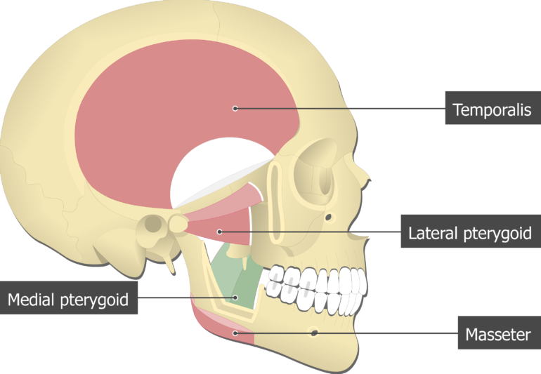
Second unilateral contraction of the medial pterygoid muscle with lateral pterygoid muscle ipsilaterally results in lateral movement of the. The medial pterygoid acts together with masseter to elevate the mandible. The medial pterygoid muscle has a triple function.
Actions of Medial Pterygoid Muscle on the mandible. Learn about the origin insertion functions and innervation of the medial pter. This muscle functions to move the lower jaw forward down and side.
It also assists the lateral pterygoid muscle with side to side mandibular motion to help with the grinding of food. It belongs to the group of masticatory muscles along with the lateral pterygoid masseter and temporal muscles. Although having different origins both heads insert on the inner.
Use your time efficiently and maximize your retention of key facts and definitions with study sets created by other students studying Medial Pterygoid Muscle Action. The medial pterygoid muscle attaches to the angle of the mandible and to the lateral pterygoid plate to form a sling with the masseter muscle that suspends the mandible Figure 6-19. They also protect the contents of the abdomen against injury and help support the body.
Where are the Pterygoids. They insert into the medial surface of the angle of the mandible posterior to the mylohyoid groove. The medial pterygoid muscle is one of the four paired muscles of mastication.
The pterygoid muscles are the two jaw. The medial pterygoid muscle attaches to the angle of the mandible and to the lateral pterygoid plate to form a sling with the masseter muscle that suspends the mandible Figure 6-19. The superficial head is attached to the maxillary tuberosity and the pyramidal process of the palatine bone.
The primary action is to elevate the mandible and laterally deviate it to the opposite side. Specifically the medial pterygoid muscle functions to. The lateral pterygoid Latin.
The medial pterygoid muscle is innervated by the medial pterygoid branch of the mandibular division of the trigeminal nerve CN V3. Medial Pterygoid OriginSuperficial head. Muscles are responsible for locomotion and play an important part in performing vital body functions.
They move the mandible side to side. Medial pterygoid has several actions. From tuberosity of maxilla adjoining boneDeep head.
It receives blood supply from the pterygoid branches of the maxillary artery. The bilateral contraction of this muscle elevates the mandible and closes the mouth. The action of the muscle during bilateral contraction of the entire muscle is to elevate the mandible raising the lower jaw.

Medial Pterygoid Muscle Attachments Actions Innervation
![]()
Medial Pterygoid Origin Insertion Action Innervation Kenhub

Medial Pterygoid Muscle Wikipedia
Medial Pterygoid Muscle Attachments Actions Innervation

Image Result For Pterygoid Muscle Human Anatomy And Physiology Muscle Anatomy Muscular System
![]()
Medial And Lateral Pterygoid Muscle Anatomy And Function Kenhub

Figure Medial Pterygoid Muscle Image Courtesy O Chaigasame Statpearls Ncbi Bookshelf
Medial Pterygoid Muscle Attachments Actions Innervation
0 comments
Post a Comment